In this week’s blog we are going to explain a section of the ADVANCE Study day in a bit more detail to help explain what participants experience when they visit us. In this post we explain why we do x-rays.
What is an X-ray?
An x-ray is a form if high-energy electromagnetic radiation that can pass through your body. An x-ray image is a black and white image created on a computerised monitor. A special machine in the x-ray room transmits a small amount of radiation which then passes through the body. The image is captured on a special image capturing device (often referred to as a digital cassette) to produce the final diagnostic image.
What are x-rays used for?
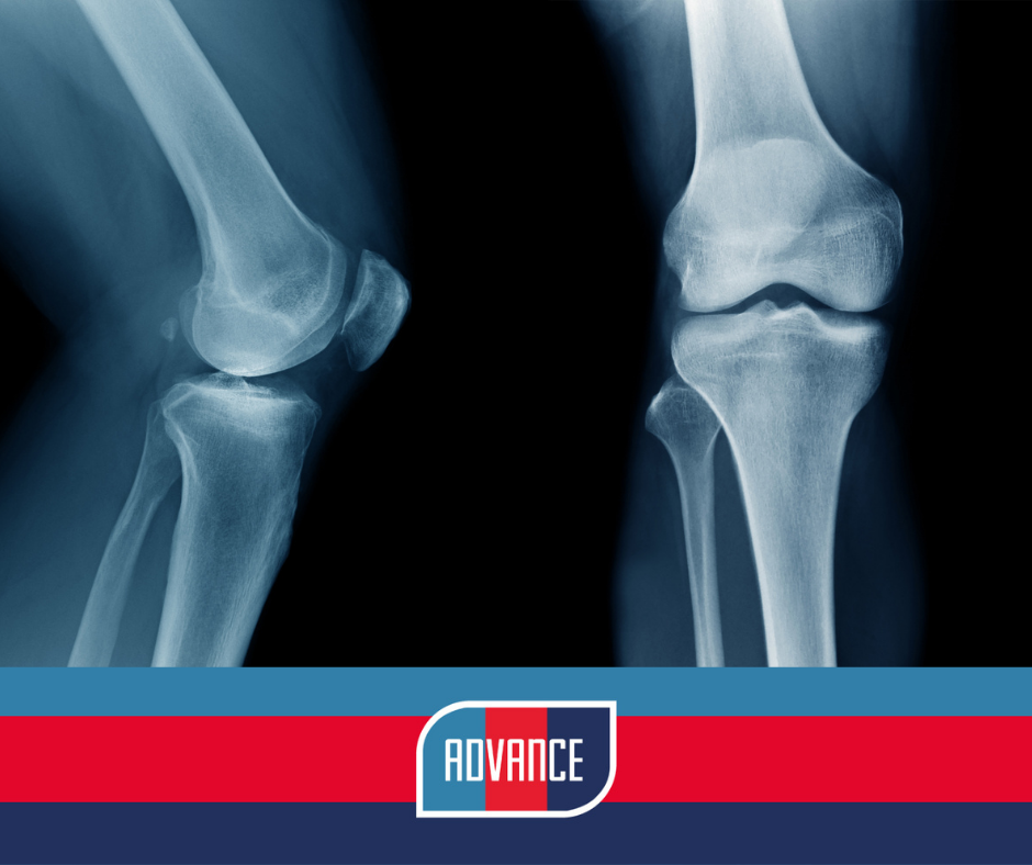
X-rays are mainly used to look at the bones, joints and surrounding soft tissue areas. X-ray examination helps diagnose and monitor conditions and injuries. During the ADVANCE visit we take x-rays of the knee and hip at various angles to look at osteoarthritis (OA).
Why do we take x-rays of the knees and hips?
Investigating osteoarthritis (OA) of the hip and knee in ADVANCE participants is one of the main aims of the study. We are really interested in whether our injured group have differences in how healthy their joints are compared to the uninjured group. Using a variety of measures we quantify the extent of wear and tear, such as how narrow the space between bones is, if there are any excessive bony overgrowths (osteophytes) within the joint space, or signs of excessive bone hardening (sclerosis). We also assess signs of wear and tear in different parts of the joint, for example, the inside and the outside of the knee joint). These factors are ranked from 0 to 4; a score of 0 is an absence of any OA changes, and a score of 4 demonstrates large osteophytes, marked joint space narrowing, severe sclerosis and abnormality of bone ends.
OA has not been looked at in a cohort of this size before, making ADVANCE very novel and teaching us a lot about the effects of trauma on joint health. We know from previous research that people who have had limb amputations have a higher rate of OA on their unamputated side compared to non-amputee controls, but we are unsure if other injuries sustained in our group may also increase the risk of OA. We also know sporting injuries like anterior cruciate knee ligament injuries increase OA risk in athletes, so it will be important for us to see if blast injuries, gunshot injuries etc. will have a similar effect on joint health.
Joint health and function
The analysis of the x-ray images will be used alongside many of the functional tests completed during ADVANCE testing and questionnaires that specifically ask about knee and hip health, pain, and function. We can then see how well the x-ray images predict function in our cohort. Ultimately, this new information should improve our knowledge of the consequences of battlefield injuries on joint health and hopefully help us improve prevention and rehabilitation of joint problems following similar trauma.